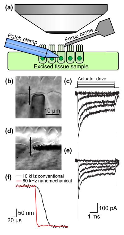
Figure 4.
Comparison between conventional and nanomechanical force probes for the study of cellular mechanotransduction. (a) Experimental setup (not to scale). (b, d) Optical micrographs of the probes in the vicinity of inner hair cells from the mammalian cochlea with the movement direction indicated. (c, e) The mechanotransducer currents evoked by a step displacement are similar and adapt with similar kinetics for a constant displacement of up to 500 nm. (f) Probe step response comparison. The nanomechanical probe is nearly an order of magnitude faster than its macroscale glass counterpart.

