
Figure 3.
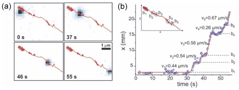
Tracking of kinesin by quantum-dot labels (Courty et al., 2006b). (a) Tracking of a quantum-dot-labeled kinesin inside a living cell. Images at different time points are shown with the overall trajectory overlaid in red. (b) Inset: trajectory of the kinesin shown in (a). The position along x axis vs time demonstrates successive directed movements (with corresponding velocities vi) and diffusive motions (marked by bi).