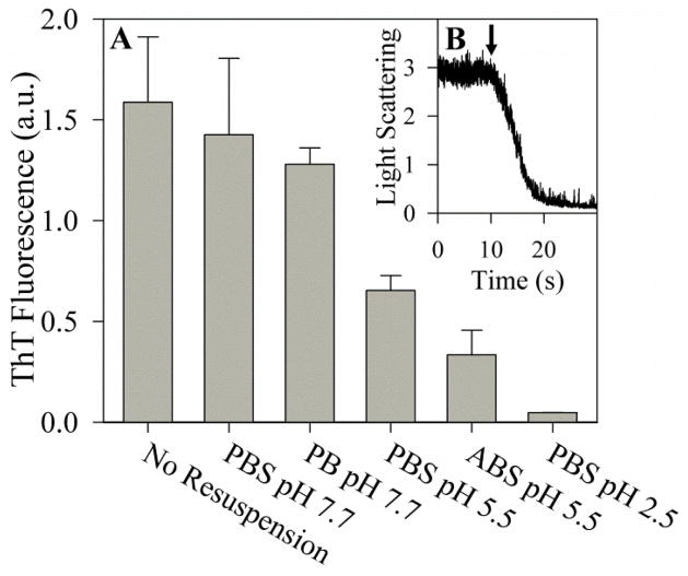
Figure 2.
Effect of pH on PAPf39 fibril dissociation. Panel A: ThT fluorescence intensity of preformed PAPf39 fibrils after 24 hours at room temperature in different buffers. Corresponding AFM images can be found in Figure S2. Panel B: Kinetics of pH-induced dissociation of preformed PAPf39 fibrils as monitored by light scattering at pH 2.8. The time point of HCl addition is indicated by an arrow.

