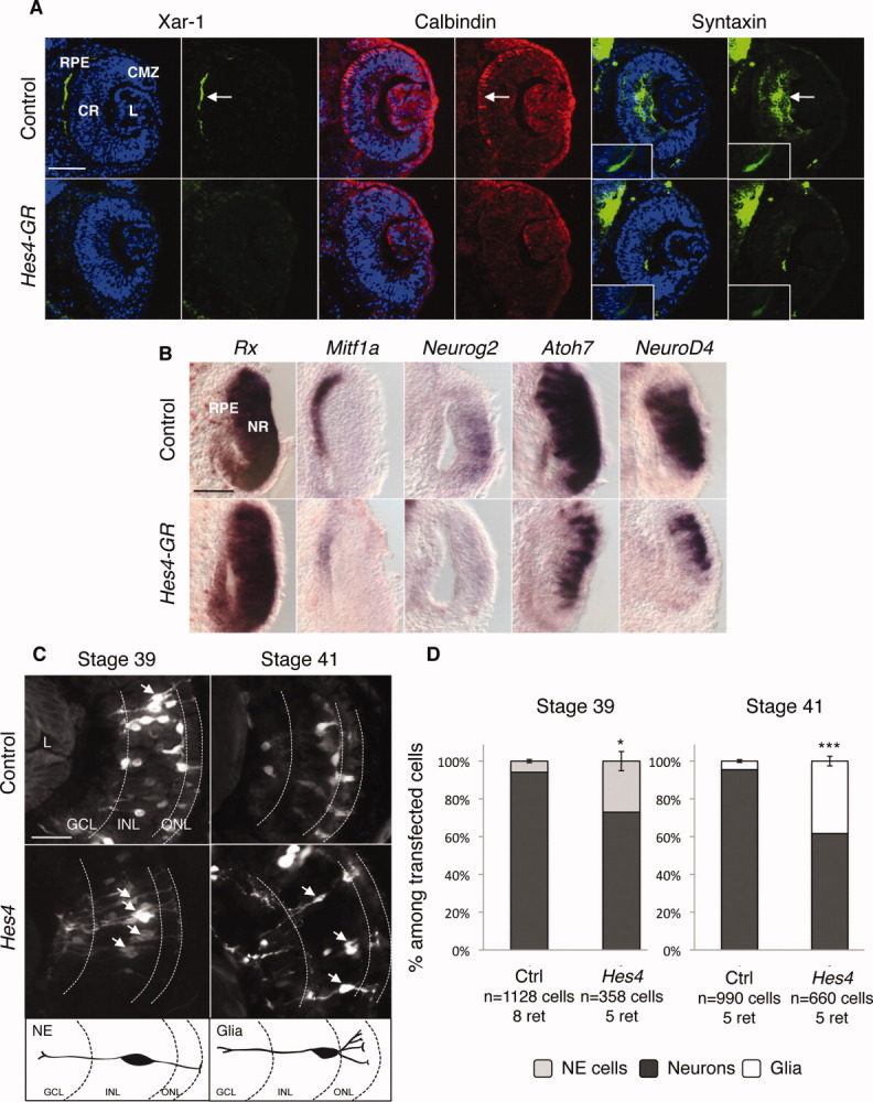
Figure 4.

Hes4 misexpression inhibits neuronal and RPE differentiation. (A): Immunofluorescence analysis of cell-type-specific marker expression in stage 37 retinas, following Hes4-GR mRNA injection. Arrows point to the staining in the RPE (Xar-1), photoreceptors (calbindin) or interneuron and ganglion cell fibers (syntaxin). The optic nerve is shown in insets. (B):In situ hybridization analysis with the indicated probe in stage 25 retinas, following Hes4-GR mRNA injection. (C, D): Analysis of cell type distribution in stage 39 or 41 retinas, following Hes4 lipofection. (C) Typical sections of retinas transfected with GFP alone (control) or GFP plus Hes4. Arrows indicate NE at stage 39 or Müller glial cells at stage 41. Respective morphologies of these cells are illustrated on the schematics below. (D) Quantification of NE, neurons, and glia among transfected cells at stage 39 or 41. Scale bars = 50 μm (A, B) or 25 μm (C). Abbreviations: CMZ, ciliary marginal zone; CR, central retina; GR, glucocorticoid receptor; GCL, ganglion cell layer; INL/ONL, inner/outer nuclear layer; L, lens; NE, neuroepithelial cell; NR, neural retina; RPE, retinal pigmented epithelium.
