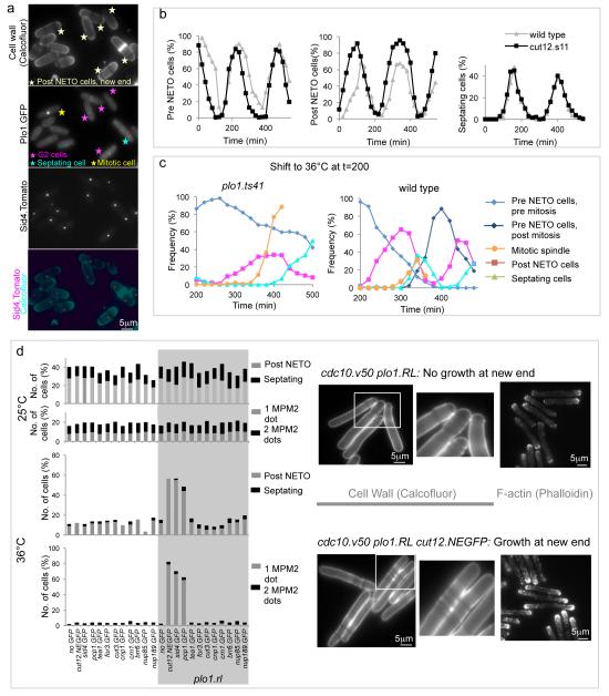
Figure 3. NETO is triggered by Plo1 activation/recruitment on the G2 SPB.
a) Fluorescence imaging of cell wall signals (calcofluor, top), Plo1.GFP and Sid4.Tom of living cells of the indicated strains. Arrows identify cells with Plo1.GFP signals on SPBs. b) Size selected small G2 cells were fixed and scored for NETO status and septation index as they transited the cell division cycle. c) Cultures in which cell cycle progression had been synchronized by the selection of small cells at t=0 were allowed to transit one cell cycle (Supplementary Figure 1g) before the temperature was shifted to 36°C and the frequency of NETO and mitotic spindle index scored in fixed cells. As indicated in Figure 1g, inactivation of Plo1 delayed mitotic commitment in the plo1.ts41 panel. Despite this delay, over 60% of cells failed to undergo NETO by the time the mitotic index rose sharply as cells accumulate with monopolar spindles (Supplementary Figure 1f). d) Mutants harbouring cdc10.v50 and the indicated GFP fusion genes were grown to early log phase at 25°C before the plo1.RL allele was induced by removal of thiamine in the presence of 40 μM 3MB-PP1. After dilution and a further 24 hours at 25°C the early log phase cells were filtered into analogue and thiamine free medium and split into two, one half being kept at 25°C, the other being shifted to 36°C (lower). 5 hours later cells were fixed and the frequency of NETO scored (left). The panels on the right show examples of the wild type control (upper) and cut12.NEGFP (lower) cdc10.v50 strains after incubation at 36°C stained with Calcofluor to highlight cell wall material or TRITC phalloidin to stain F-actin.

