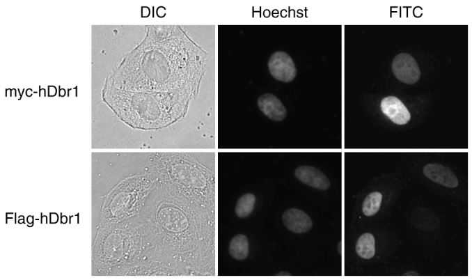
Figure 2. hDbr1 localizes to the nucleoplasm by immunofluorescence.

Expression vectors encoding either myc-tagged (upper panels) or Flag-tagged hDbr1 (lower panels) were transfected into HeLa cells. Twenty-four hours after transfection, the cells were stained with either anti-myc (MC045, Nacalai Tesque, Japan, panel FITC, upper right) or anti-Flag (M2, SIGMA, panel FITC, lower right). The cells were also stained with Hoechst 33342 (SIGMA, panel Hoechst, middle) to label the nuclei. Differential interference contrast (DIC) images of the cells are also shown as DIC panels at the left.
