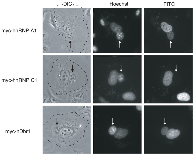
Figure 5. The hDbr1 protein shuttles between the nucleus and the cytoplasm.
Expression vectors encoding hnRNP A1, C1, and hDbr1 with myc tag were transfected into HeLa cells. After expression of the transfected cDNAs, the cells were fused with mouse NIH3T3 cells to form heterokaryones and incubated in media containing 100 μg/ml cycloheximide for 2 hours. The cells were then fixed and stained for immunofluorescence microscopy with anti-myc tag antibody (panel FITC) to localize the proteins, and Hoechst 33342 (SIGMA, panel Hoechst), which differentiates the human and mouse nuclei within the heterokaryon. The arrows identify the mouse nuclei. The panels marked DIC show the phase-contrast image of the heterokaryons and the cytoplasmic edge is highlighted by a broken line.

