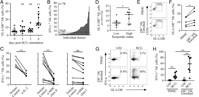
Figure 5.

Enrichment of HLA-DR+ NK cells enhances the IFN-γ response to BCG. (A) PBMCs from ten donors were cultured with BCG for up to 6 days and the percentage of CD56+CD3− NK cells staining positive for HLA-DR was measured by flow cytometry, median±interquartile range, **p<0.01 tested by Wilcoxon matched pairs. (B) PBMCs from 78 donors were cultured with BCG for 24 h then analysed by flow cytometry for intracellular IFN-γ in NK cells, dotted line represents the median (6.5%). (C) PBMCs from ten donors were cultured with BCG for 24 h with blocking antibodies to MHC molecules or to IL-2 or with isotype matched controls. The percentage of IFN-γ+ NK cells was determined by flow cytometry, **p<0.01 tested by Wilcoxon matched pairs. (D) PBMCs from 16 donors (seven high and nine low responders) were analysed by flow cytometry for the percentage of HLA-DR-expressing NK cells, median±interquartile range, *p<0.05 tested by Mann Whitney one-tailed test. (E) PBMCs were enriched for HLA-DR+ NK cells and confirmed by flow cytometry; gates indicate percentage of HLA-DR+ NK cells. (F) HLA-DR-expressing NK cell enrichment for five donors, *p<0.05 tested by Mann Whitney. (G) PBMCs (top) or HLA-DR+ NK cell-enriched PBMCs (bottom) were cultured for 24 h with growth medium control (GM, left) or with BCG (right) then analysed by flow cytometry for intracellular IFN-γ in CD56+CD3− NK cells; gates indicate percentage of IFN-γ+ NK cells. (H) Data for five donors, median±interquartile range, **p<0.01 tested by Wilcoxon matched pairs.
