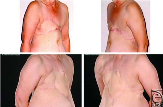
Figure 5.
Case 3: Further local recurrence and reconstruction, including advancement of the reverse abdominoplasty. Recurrent disease, visible as a 6 × 8 × 3 cm epigastric bulge above the umbilicus (top row), was radically resected and reconstructed with a left latissimus dorsi flap and advancement of the reverse abdominoplasty flap. Appearances 2 years after surgery (bottom row) show no local recurrence and an acceptable aesthetic appearance of the 3-flap reconstruction.

