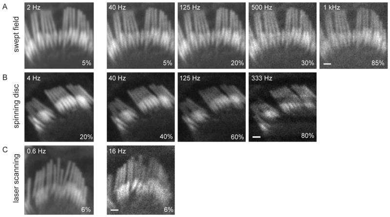
Figure 3.
Comparison of different sampling rates in three confocal systems. A: A hair bundle was imaged with the SFC system at different acquisition rates (2 to 1,000 fps) using a 50 μm slit. The laser intensity, depicted in the lower right corner of each panel, had to be increased for higher frequencies to obtain equivalent illumination intensities. B: Another hair bundle was imaged with the CSU10 Yokogawa spinning disc system at different sampling rates (4 to 333 fps) using a 50 μm pinhole. The maximal rate in our spinning disc microscope was 360 fps. Scan line inhomogeneity observed at higher rates is due to the inability of our spinning disc head to change its rotation speed to have integer scans of the image during our camera exposure time. C: Another hair bundle was imaged with the LSM 5 Exciter laser scanning confocal system at two acquisition rates. The image was set to 80 × 80 pixels for the sake of comparison. The fastest frequency allowed by the LSCM was 16 fps using 4 μs dwelling time. Scale = 1 μm.

