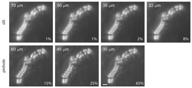
Figure 4.

Comparison of a hair bundle SFC section imaged with different slits and pinhole sizes. Rat hair cell bundle was fixed and stained with phallodin-Alexa Fluor 488. Images were acquired at 40 fps during 500 ms using different slit or pinhole sizes. Slit widths closer to the Airy limit improved resolution. Contrast was improved by using pinholes to the detriment of light exposure. The laser intensity is depicted in the lower right corner of each panel. Scale = 1 μm.
