
Figure 4.
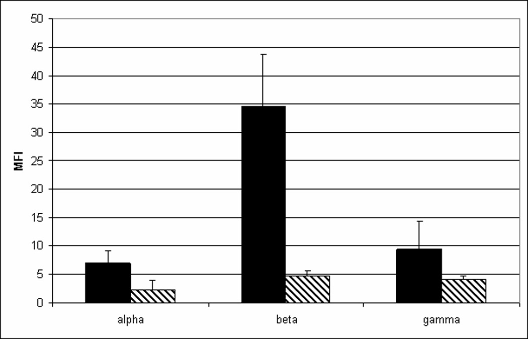
TFPIγ produced by CHO cells is secreted and inhibits TF-fVIIa activity. A) CHO cells were transfected with mouse TFPIα, TFPIβ or TFPIγ and examined for surface TFPI expression using flow cytometry following incubation for 30 min at 37°C in buffer (solid bars) or in the presence of 1 U/ml PIPLC (hatched bars). B) The amount of TFPIγ activity in conditioned media (16 h culture) and the PIPLC releasate of CHO cells transfected with TFPIγ was determined in assays that measure in TF-fVIIa activation of fX: (■) Buffer only; (♦) Conditioned media; (▲) PIPLC releasate; (●) Conditioned media in the presence of inhibitory anti-mouse TFPI antibody; (▼) PIPLC releasate in the presence of inhibitory anti-mouse TFPI antibody. C) TFPI activity was quantified in conditioned media (16 h culture) and the PIPLC releasate using assays that measure in TF-fVIIa activation of fX. The concentration of TFPI in the media and PIPLC releasate was determined by comparison to a standard curve generated using known amounts of recombinant TFPI and multiplied by the sample volume to obtain the total amount of TFPI in each sample. Data are expressed as the percentage of the TFPI released from the cell surface by PIPLC (average ± SD of three experiments).