
Figure 5.
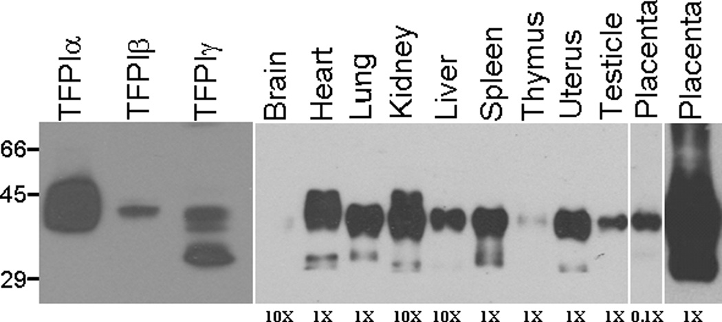
Western blot analysis of TFPI in mouse tissues. Mouse tissues were lysed in CHAPS buffer, protein standardized and subjected to SDS-PAGE and western blot analysis for TFPI. The relative amount of total protein from each tissue loaded on the gel is indicated at the bottom of the gel. TFPIα, TFPIβ and TFPIγ from transfected CHO cells were also subjected to SDS-PAGE and western blot analysis for comparison with the tissue blots.