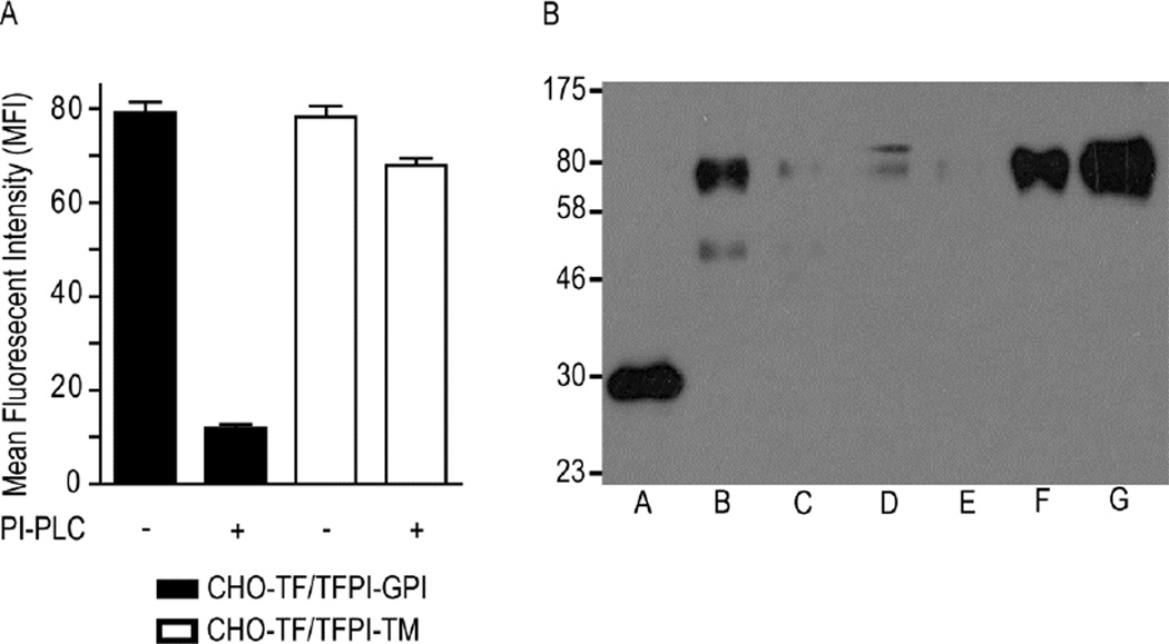
Figure 2. TFPI-GPI is localized to caveolae, while TFPI-TM is localized to the bulk plasma membrane.
A) Flow cytometric analysis of TFPI expression on CHO-TF/TFPI-GPI and CHO-TF/TFPI-TM cells before and after PI-PLC treatment; mean ± SD, n=4. B) Triton X-114 phase separation was performed on CHO-TF/TFPI-TM (Lanes B–D) and CHO-TF/TFPI-GPI cells (Lanes E–G), and the fractions analyzed for TFPI expression by Western blotting. Lane A: recombinant TFPI (32 kDa), expressed in E. coli and run as an antibody positive control; Lanes B and E: TX-114 aqueous phase; Lanes C and F: TX-114 detergent phase; Lanes D and G: TX-114 detergent insoluble pellet. The chimeras migrate to approximately 70 kDa, similar to that reported previously [17]. The lower band in lane B is a possible degradation product, while the upper band in lane D may be dimerization of the chimera [17] or a non-specific product.

