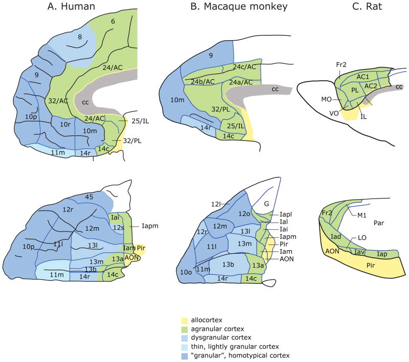
Figure 1.
Architectonic maps of the medial (top) and orbital (bottom) surfaces of the frontal lobe in A) humans 89 and B) monkeys 39. C) Medial (top) and lateral (bottom) frontal cortex in rats 90. Agranular cortex lacks layer IV. Dysgranular cortex contains a rudimentary layer IV. Granular cortex has a well-developed layer IV. Layer IV neurons are described as granular because their cell bodies are small and round and changes in this layer are clearly visible as one transitions from agranular to granular cortex. Abbreviations: AON, anterior olfactory nucleus; Fr2, second frontal area; I, insula; LO, lateral orbital area; M1, primary motor area; Par, parietal cortex; Pir, Piriform cortex; AC, anterior cingulate area; cc, corpus callosum; IL, infralimbic cortex; MO, medial orbital area; PL, prelimbic cortex; VO, ventral orbital area; l, lateral; m, medial; o, orbital; r, rostral; c, caudal; i, inferior; p, posterior; s, sulcal; v, ventral. Numbers indicate cortical fields, except that after certain areas, such as Fr2 and AC1, they indicate subdivisions of cortical fields. a has two meanings: in Ia, it means agranular; in 13a, it distinguishes that area from area 13b. Figures reproduced with permission 91.

