
Figure 6. Fbxw7 targets c-Myc for degradation during ES cell differentiation.
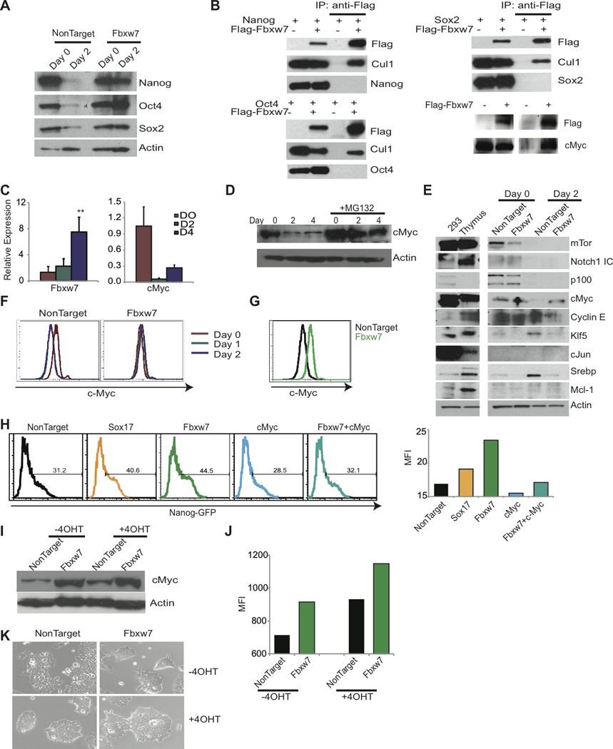
A) Western blot of pluripotency factors following Fbxw7 silencing in Day 0 and 2 of differentiation. B) Co-transfection of Fbxw7 and pluripotency factors in 293T cells followed by immunoprecipitation. Lane 1 and 2; whole cell extract and Lane 3 and 4; immunoprecipitates. C) qRT-PCR of Fbxw7 and c-Myc during differentiation. D) c-Myc protein expression during differentiation with or without 10µM MG132 treatment. E) Immunoblot of known Fbxw7 substrates at Day 0 and Day 2 of differentiation. F and G) Intracellular FACS staining with anti-c-Myc following shRNA transduction and differentiation with RA. H) Histogram representing retention of GFP 72hrs following siRNA transfection and 48hrs addition of differentiation media. Bar graph on the right represents MFI of ES cells in one representative experiment (n=3). I-J) MycT58AER ES cells following depletion of Fbxw7 with shRNA were differentiated for 2 days (−LIF+5µM RA) without or in the presence of 10nM 4-OHT, and analyzed by I) immunoblot for c-Myc, J) SSEA1 expression Bar graph represents MFI of SSEA1 in one representative experiment (n=2), and K) morphology of colonies. Data represented as +SEM; N=3. *p-value <0.05, **p-value<0.01.