
Figure 7. Psmd14 and Fbxw7 regulate cellular reprogramming.
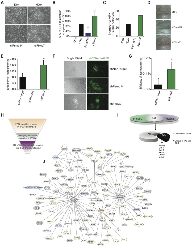
A) Images of ubiquitously expressed actin-RFP 6 days post siRNA transfection and Dox (OKSM) induction demonstrating morphology changes. B) AP+ ES-like colony count at Day 14 following siRNA knockdown of Fbxw7 and Psmd14 relative to +Dox control. C) Absolute number of AP+ ES-like colonies at day 14 following siRNA transfection and addition of Dox of MEFs. One representative experiment is shown (n=4). D) AP staining 14 days following siRNA transfection and addition of Dox. E) Reprogramming efficiency of OKSM MEFs expressing shRNAs against non-target, Psmd14, or Fbxw7 to generate AP+ ES-like colonies. Colonies were enumerated at day 14. F) Morphology of representative colonies following transduction of shRNAmir at day 14. Green; shRNAmir transduced cells. G) Efficiency of OSK MEFs expressing shRNAs against NonTarget, or Fbxw7 to generate AP+ ES-like colonies. Colonies were enumerated at day 14. H) Proteins identified and quantified in MEFs and iPS in previous studies, showing proteins highly expressed in iPS and the number of the ones that are ubiquitinated. I) Overlap between ubiquitinated proteins in ES and iPS and comparison with ubiquitinated proteins in MEFs. J) Self-Renewal network, as depicted in Figure 1D, demonstrating proteins that have been identified to be ubiquitinated in iPS and ES (grey). Proteins also ubiquitinated in MEFs are marked in yellow. Proteins in orange were found ubiquitinated in iPS and MEFs and proteins in purple were exclusively found in iPS. Data represented as +SEM; N=3. *p-value <0.05, **p-value<0.01.