Figure 3.
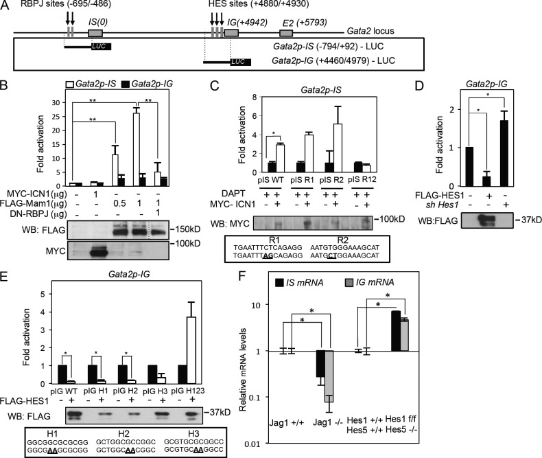
HES-1 is a negative regulator of the Gata2 promoter in hematopoietic cells. (A) Scheme of the Gata2 locus indicating the conserved RBPJ- and HES-binding sites (vertical arrows). Promoter regions were used in the luciferase assay. (B) Luciferase activity from the Gata2p-IS and Gata2p-IG promoter in the presence or absence of ICN1, Mam1, or dn-RBP in HEK-293T cells (**, P ≤ 0.009). The dotted line in the Western blot (WB) indicates that intervening lanes have been spliced out. (C) Luciferase activity from the Gata2-IS promoter with indicated point mutations in the RBPJ-binding consensus (bold/underlined) measured in HEK-293T cells. DAPT incubation was used to reduce endogenous Notch activity. (D) Luciferase activity of the Gata2-IG promoter in the presence or absence of HES-1. (E) Luciferase activity from the Gata2-IG promoter with the indicated point mutations (bold/underlined) in the HES-binding consensus. (B–E) Mean ± SD of at least three independent experiments is shown in all the plots; Student’s t test was used to assess the significance (*, P < 0.05). (F) Relative expression of the indicated genes from independent determinations of fresh E10.5 AGMs in Jagged1+/+ (n = 3) and Jagged1−/− (n = 3) and E10.5 AGMs in Hes1-5 wild type (n = 2) and knockouts (n = 3) by qRT-PCR after 3 d of 4-OH-tamoxifen treatments in explant cultures. Fold increase to the wild-type control after GAPDH normalization is shown. Bars represent the mean ± SD determination; Student’s t test was used to assess the significance (*, P < 0.05).

