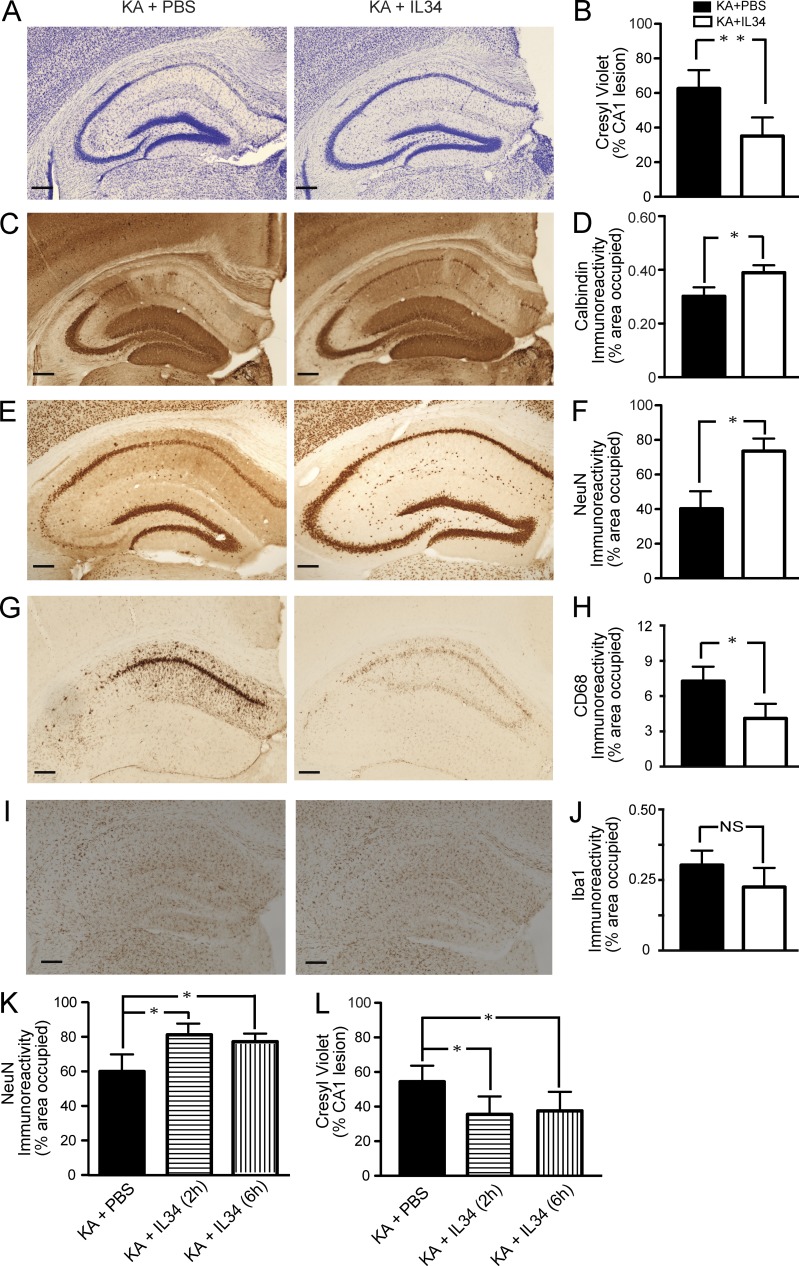
Figure 4.
Systemically administered IL-34 attenuates KA-induced neurodegeneration and microgliosis. (A–J) 2-mo-old FVB/N mice were lesioned with 20 mg/kg KA (subcutaneous injection) and sacrificed 5 d later. 100 µg/kg IL-34 was injected i.p. once 2 h before KA. KA-induced neuronal injury was assessed by cresyl violet staining (A and B), calbindin (C and D), and NeuN (E and F) immunostaining, and microglial activation was assessed by CD68 (G and H) or Iba-1 (I and J) immunostaining. Representative images from two independent experiments are shown from hippocampi of mice treated with PBS (left) or IL-34 (right). Bars, 200 µm. Bars in B, D, F, H, and J are mean ± SEM (n = 4 mice/group) from one out of two independent experiments. *, P < 0.05; **, P < 0.01 compared by Student’s t test. (K and L) KA-lesioned mice were treated with IL-34 at 2 and 6 h after injury. The mice were sacrificed at day 5. Excitotoxic injury was assessed by NeuN immunostaining (K) and cresyl violet staining (L). The experiment was performed once. Bars are mean ± SEM (n = 5 mice/group). *, P < 0.05 compared by ANOVA and Bonferroni post-hoc test.

