Figure 6.
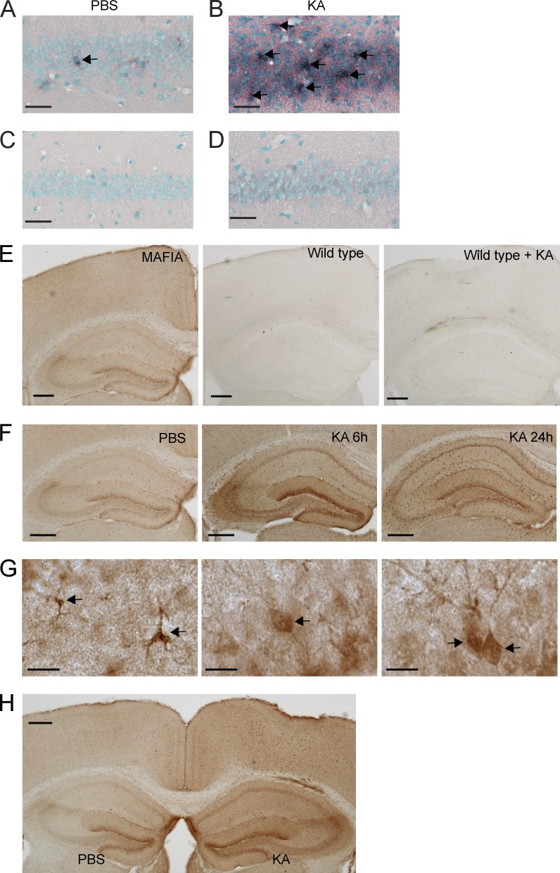
Expression of CSF1R in neurons. (A–D) Detection of Csf1r mRNA in the hippocampus using in situ hybridization. Wild-type FVB/N mice were lesioned with 20 mg/kg KA (subcutaneous injection) or injected with PBS as a control and sacrificed 24 h later. Brain sections were hybridized with DIG-labeled antisense (A and B) or sense (C and D) probes for Csf1r and developed with an anti-DIG antibody. No signal was detected with the sense probe (C and D). Arrows point to cells expressing CSF1R, as detected by in situ hybridization. (E–H) CSF1R reporter mice (MAFIA mice, 2 mo of age) were lesioned with 20 mg/kg KA (subcutaneous injection) or injected with PBS as control and sacrificed 6 and 24 h later. These mice express an EGFP tag under the control of the Csf1r promoter so that the expression of CSF1R can be detected by GFP immunostaining. (E) To test the specificity of the antibody against GFP, brain sections from MAFIA mice (not injured, left), wild-type mice (middle), and wild-type mice with KA injury (right) were immunostained. (F) Representative images of different brain regions from MAFIA mice injected with PBS and sacrificed 24 h later (left) or injected with KA and sacrificed at 6 (middle) or 24 (right). (G) High-magnification images showing GFP immunoreactivity in microglia (left) and neurons (middle and right), determined by morphology. Arrows point to cells expressing CSF1R, as detected by immunohistochemistry for the reporter gene. (H) 2-mo-old MAFIA mice received unilateral stereotaxic injections of 50 ng KA (right hemibrain) or PBS (left hemibrain). Mice were sacrificed 6 h later, and brain sections were analyzed for reporter gene expression by GFP immunostaining. The data are representative of two independent experiments with n = 3 mice/group. Bars: (A–D) 50 µm; (E, F, and H) 200 µm; (G) 10 µm.

