Figure 7.
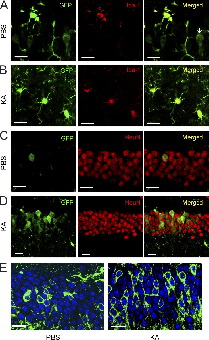
CSF1R reporter gene is expressed in neurons and up-regulated after excitotoxic brain injury. (A–D) CSF1R reporter mice (MAFIA mice, 2 mo of age) were lesioned with 20 mg/kg KA (subcutaneous injection) or injected with PBS as control and sacrificed 6 h later. Representative confocal microscopy images from control (PBS; A and C) and KA-lesioned mice (B and D) double immunolabeled with antibodies against GFP and cell type–specific markers Iba-1 (microglia; A and B) and NeuN (neurons; C and D). The reporter gene–expressing cells appear yellow after superimposition. An Iba-1 immuno-negative cell that expresses the reporter gene is shown (arrow) in A. Up-regulation of the reporter gene in neurons is shown after KA lesion (D) compared with control (C). The data are representative of two independent experiments with n = 3 mice/group. (E) Csf1r-iCre mice were crossed with mTmG mice. Cre recombinase expression driven by the endogenous Csf1r promoter leads to expression of EGFP in double transgenic mice. Representative confocal microscopy images from control (PBS; left) and KA-lesioned mice (right) 6 h after injury double immunolabeled with antibodies against GFP (green) and neuron-specific marker NeuN (blue). This experiment was performed once with n = 5 mice/group. Bars, 20 µm.

