Figure 8.
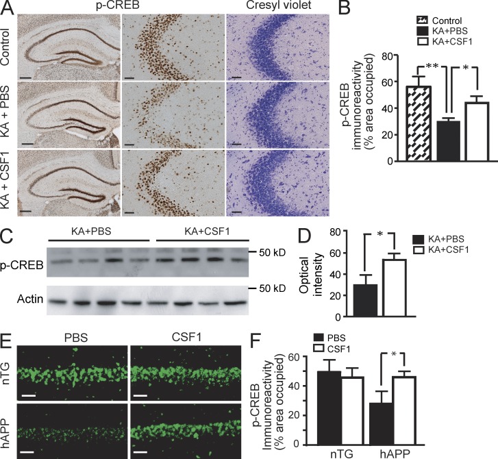
CSF1 maintains CREB phosphorylation. (A–D) CSF1 maintains CREB phosphorylation after KA lesion. Wild-type FVB/N mice (n = 4 per group, 2 mo of age) were lesioned with 20 mg/kg KA (subcutaneous injection) or injected with PBS as control. For treatment, CSF1 or PBS was applied 24 h before KA. Mice were sacrificed 6 h after KA injection. One hemibrain was fixed for immunohistochemistry with an antibody against p-CREB (A, left and middle), and p-CREB immunoreactivity was quantified as percentage of area occupied (B). The opposite hippocampi were isolated and subjected to Western blot analysis for p-CREB (C), which was quantified as optical intensity (D). Results are from one out of two independent experiments. (E and F) CSF1 restores CREB signaling in hAPP mice. hAPP mice and their nTG littermates (Fig. 1 A) were treated with 800 µg/kg CSF1 or PBS three times a week. 10 wk later, the mice were sacrificed after water maze behavior test, and one hemibrain was analyzed by immunohistochemistry for p-CREB immunoreactivity. The experiment was performed once. Error bars indicate SEM. Bars: (A, left) 200 µm; (A [middle and right] and E) 50 µm. *, P < 0.05; **, P < 0.01 compared by ANOVA and Bonferroni post-hoc test (B and F) or Student’s t test (D).

