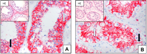
Figure 1.

N-cadherin expression in normal testis and IGCNU: Strong cytoplasmic (white arrow) and a weak membranous (black arrow) expression is seen in the normal testis (A; x200). In representative tissues of IGCNU, a more pronounced membranous expression (black arrow) and a cytoplasmic (white arrow) is seen (B; x200). N-cadherin is not detectable in sperms and cells of the interstitium, including Leydig cells (A - B).
