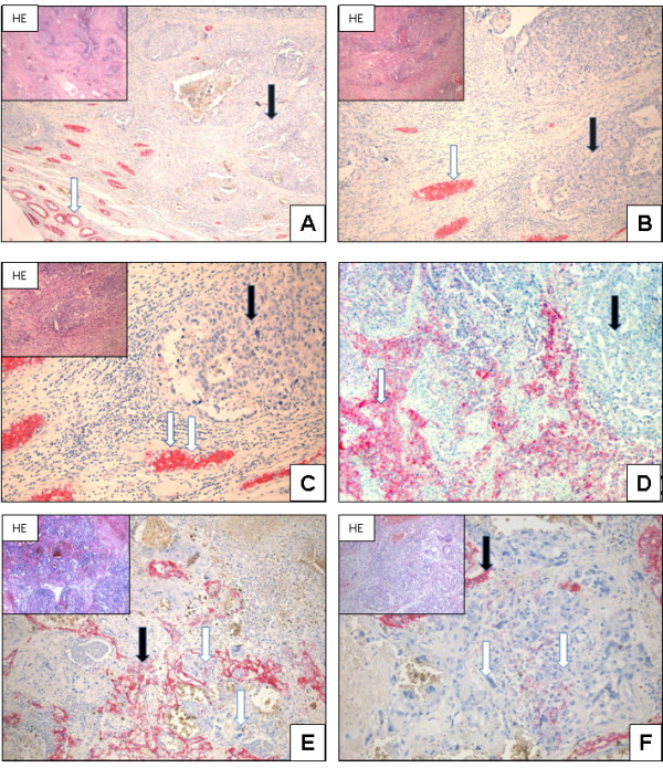
Figure 4.

N-cadherin expression in embryonal carcinoma and chorionic carcinoma: Embryonal carcinoma: Normal testis and IGCNU showing N-cadherin expression (white arrow). In contrast, the embryonal carcinoma cells do not show any expression of N-cadherin (black arrows, A; x40, B; x200, C; x400). Even mixed tumours with embryonal carcinoma are negative for N-cadherin (black arrow), whereas seminoma shows N-cadherin expression (white arrow, D; x100). Chorionic carcinoma: Tumour cells and syncytiotrophoblastic cells do not show N-cadherin expression (white arrow). Adjacent tumour cells (yolk sac tumour components) are positive for N-cadherin expression and show membranous and cytoplasmic staining (black arrow, E; x40, F; x200).
