
Figure 3.
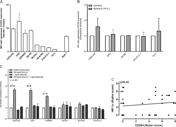
Expression and functionality of CXCR4 in Ewing sarcoma: role for CXCL12-dependent modulation of tumor cell proliferation. A. Constitutive surface expression of CXCR4 in EWS cell lines, as assessed by flow cytometry. Results are expressed as the mean ± SD MFI-ratio, obtained in at least two independent experiments. Breast cancer cell line MCF-7 was used as a positive control. B. Flow cytometric analysis of 24-hours hypoxia (0.1% O2)-induced CXCR4 expression in cell lines having either substantial (CADO-ES, EW3, RD-ES) or minimal (STA-ET2.1, TC71) levels of constitutive CXCR4 expression. Results are expressed as the mean ± SD fold increase in MFI-ratio over normoxic control, obtained in at least two independent experiments. Culture under hypoxic conditions (as previously described [18]) did not systematically affect CXCR4 expression. C. Pearson correlation analysis: lack of correlation between in vivo HIF-1α [18] and CXCR4 protein expression levels in 38 therapy-naïve EWS. D. Stimulation of cell lines expressing substantial levels of CXCR4 (CADO-ES, EW3, IOR/BER) with 100ng/ml recombinant CXCL12 for seven days significantly increased cell numbers. Addition of AMD3100 (1μg/ml) abrogated the increase in cell numbers. No effects were observed in cell lines with minimal levels of CXCR4 expression (SK-ES-1, SK-N-MC, STA-ET2.1), nor did AMD3100 treatment alone affect cell proliferation. Cells were cultured in serum-free medium. Cell viability was measured by MTS cell viability assay, a colorimetric method for determining the number of viable cells in proliferation assays (see Methods section). Results are expressed as the mean ± SD fold increase in cell numbers over (untreated) medium control, obtained in at least two independent experiments.