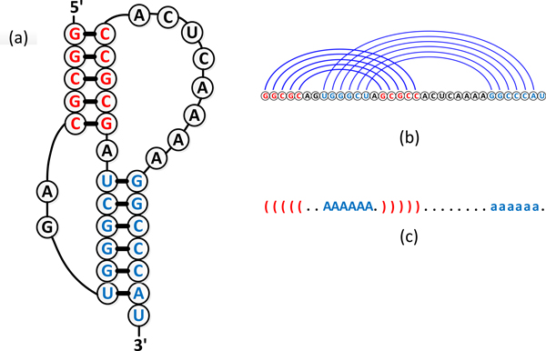
Figure 1.

Structure of an RNA pseudoknot. (a-c) show the secondary structure, arc-based representation, and dot-bracket notation of mouse mammary tumor virus (MMTV) H-type pseudoknot with PDB code 1RNK. The bases in stacking regions are colored with blue while the unpaired bases are colored with black.
