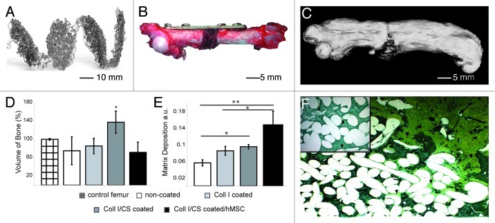
Figure 6. In vivo study small animals: created 5 mm orthotopic critical size defect (femur) in immunodeficient nude rat. Implantation of non-coated, Coll I or Coll I/CS coated, as well as Coll I/CS coated/hMSC seeded PCL scaffolds over 12 weeks, five animals per group. (A) Specially designed PCL scaffolds for rat femur critical size defects with a thickness of 0.5 mm and a diameter of 5 mm. (B) A 5-hole mini-fragment plate was fixed to the femur and a 5 mm long segmental mid-diaphysial osteotomy was performed. (C) 3-dimensional CT reconstruction of a rat femur (Coll I/CS group) showing callus formation along the femur. The new bone formed along and into the scaffold pad up to bridging the critical size defect. (D) Quantification of newly produced bone volume in the defect zone showed the highest amount of new bone in the Coll I/CS group (137%) compared with the non-coated (75%), Coll I (85%), or Coll I/CS/hMSC (72%) group. Non-operated contralateral femora were used as control (100%) (significance: *p < 0.05). (E) Quantification of the matrix deposition using a modified trichrome Masson-Goldner staining in the defect zone showing the highest matrix accumulation in the Coll I/CS/hMSC followed by the Coll I/CS group, the Coll I group and the non-coated group, a.u. arbitrary units (significances: *p < 0.05, **p < 0.01). (F) New bone formation occurred at the proximal and distal ends of the defect zone, localized around the scaffold pad and in the bordering scaffold areas (star, green coloring). The central part showed variable amounts of matrix aggregation (arrow, yellow coloring) depending on the surface modification of the scaffold (Coll I/CS/hMSC > Coll I/CS > Coll I > non-coated).10

An official website of the United States government
Here's how you know
Official websites use .gov
A
.gov website belongs to an official
government organization in the United States.
Secure .gov websites use HTTPS
A lock (
) or https:// means you've safely
connected to the .gov website. Share sensitive
information only on official, secure websites.
