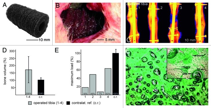
Figure 7. In vivo study large animals: created 3 cm orthotopic critical size defect (tibia) in sheep. Implantation of Coll I/CS coated PCL scaffolds over 12 and 48 weeks, five animals per group and time point. (A) Thirty piled scaffolds forming a 3-dimensional implant. (B) Three centimeter long mid-diaphysial defect in the sheep tibia filled with 30 piled scaffolds. (C) Radiological investigation (false coloring) of four sheep tibia defect areas showing two tibial defects were bridged completely after 12 mo (2 and 4). One showed a hypertrophic non-union (1) and one an atrophic non-union (3). (D) The quantification of the bone volume ratio after 12 mo shows the newly formed bone averaged 172% (+/− 86%) as compared with the intact contra lateral tibiae used as a reference value (100%). (E) Biomechanical evaluation at 12 mo (maximum load until failure) demonstrated that two operated tibiae (1 and 4) reached 49% (2,880 N) and 63% (3,720 N) of the reference value for non-operated bone respectively (c.r., 100%, 5,875 N). The values in the other two animals (2 and 3) reached 18% (1,050 N) and 7% (428 N). (F) Trichrome Masson-Goldner staining. Bone formation took place directly around the scaffold fibers revealing no interconnected gaps. The newly formed lamellar bone inside the scaffolds presented osteons including Haversian canals suggesting regular bone formation. According to their natural localization, osteocytes (ocy) and osteoblasts (obl) could be localized within the bone or at the adjacent areas. The scaffold was completely vascularized (bv) and erosion of the PCL fibers was clearly visible. No inflammatory reaction was evident around the implant material after 12 mo.

An official website of the United States government
Here's how you know
Official websites use .gov
A
.gov website belongs to an official
government organization in the United States.
Secure .gov websites use HTTPS
A lock (
) or https:// means you've safely
connected to the .gov website. Share sensitive
information only on official, secure websites.
