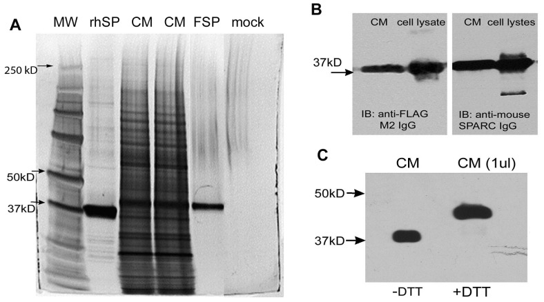
Figure 1. Production and purification of FSP.
Conditioned medium (CM) from baculovirus infected Sf21 insect cells was subjected to immuno-affinity chromatography on anti-FLAG M1 affinity gel. CM (15 ul of culture supernate), rhSP (rhSPARC, purified by anion-exchange chromatography), FSP (5 ul of eluted fraction), and mock (buffer control) were separated by SDS-PAGE on a 10% gel under non-reducing conditions, and were visualized by silver stain (A); note the other bands in the purified rhSPARC sample, but not in the FSP sample (B), 1 ul of CM of infected Sf21 cell lysates (∼5 ug total protein per lane) was resolved by SDS-PAGE under non-reducing conditions, transferred to a PVDF membrane, and probed with anti-FLAG M2 IgG or polyclonal anti-mouse SPARC IgG. (C), 1 ul CM was diluted in sample buffer and was boiled for 5 min in the presence (+) or absence (−) of 50 mM DTT. Proteins were resolved by SDS-PAGE, followed by immunoblotting for SPARC. A shift of FSP from Mr ∼38,000 to ∼44,000 was apparent.

