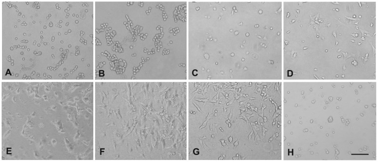
Figure 2. FSP supports mLEC attachment and spreading in a concentration-dependent manner.
(A) BSA (10 ul/ml), (B) rhSPARC (10 ug/ml), (C–G) FSP (5, 10, 20, 30, 40 ug/ml, respectively), and (H) FSP (10 ug/ml) were preincubated with anti-SPARC IgG (30 ug/ml) at 37°C for 30 min prior to the coating. The proteins were coated onto wells overnight at 4°C. mLECs were plated into each well in serum-free DMEM, and allowed to attach for 2 hr at 37°C. Phase-contrast photomicrographs were taken on the 96-well plates under an inverted microscope equipped with a digital camera. Scale bar = 50 um for all photographs.

