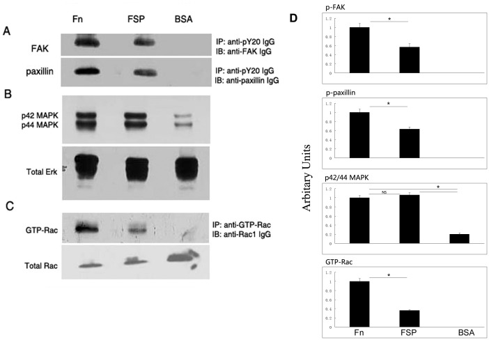
Figure 7. FSP induces phosphorylation of FAK, paxillin, and ERK1/2, and activates the small GTPase Rac in LEC.
LEC cultured in serum-free DMEM for 48 hr were plated on dishes precoated with Fn, FSP, or BSA for 1 hr at 37°C. Cell lysates were prepared. (A), immunoprecipitation of total cell lysates was performed with an anti-pY20 antibody followed by immunoblotting with anti-FAK IgG or anti-paxillin IgG. (B), Total cell lysates were immunoblotted with anti-phospho-Erk-1 (p42 MAPK) and anti- phospho-Erk-2 (p44 MAPK). Subsequently, the membrane was stripped and re-probed with anti-Erk IgG (total Erk). (C), Total cell lysates were incubated with anti-GTP-Rac IgG; activated Rac was affinity-precipitated and subsequently immunoblotted with IgG against Rac. An aliquot of cell lysate from each sample was immunoblotted for total Rac protein. (D) The histogram on the right shows results of scanning densitometry of p-FAK, p-paxillin, p42/44 MAPK, and GTP-Rac of three experiments with mean +/− SD. Data were normalized to the loading control and were plotted relative to Fn levels. *P<0.05; NS, not significant.

