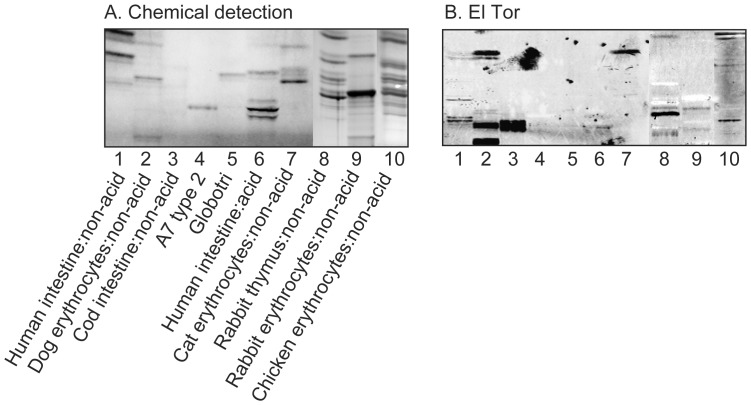
Figure 1. Binding of V. cholerae El Tor to mixtures of glycosphingolipids.
(A) Chemical detection by anisaldehyde. (B) Autoradiograms obtained by binding of V. cholerae JBK 70. The lanes were: Lane 1, non-acid glycosphingolipids of human small intestine, 20 µg; Lane 2, non-acid glycosphingolipids of dog erythrocytes, 20 µg; Lane 3, non-acid glycosphingolipids of cod intestine, 20 µg; Lane 4, A type 2 heptaosylceramide (GalNAcα3(Fucα2)Galβ4(Fucα3)GlcNAcβ3Galβ4Glcβ1Cer), 4 µg; Lane 5, globotriaosylceramide (Galα4Galβ4Glcβ1Cer), 4 µg; Lane 6, acid glycosphingolipids of human small intestine, 40 µg; Lane 7, non-acid glycosphingolipids of cat erythrocytes, 40 µg; Lane 8, non-acid glycosphingolipids of rabbit thymus, 40 µg; Lane 9, non-acid glycosphingolipids of rabbit erythrocytes, 40 µg; Lane 10, non-acid glycosphingolipids of chicken erythrocytes, 40 µg.

