
Figure 3. Subcellular localization of PAF95 and PAF96 in conidial germlings of N. crassa.
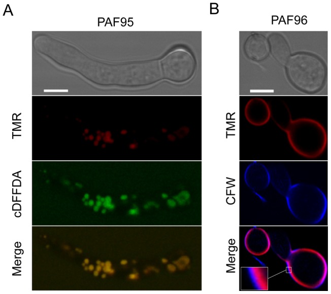
(A) Co-localization of TMR-PAF95 (in red) with the vacuolar marker cDFFDA (in green) after treatment of the germlings with 5 µM TMR-PAF95 for 1 h. Note co-localization within the vacuolar network in these merged images. (B) Localization of TMR-PAF96 (in red) and the cell wall stain CFW (in blue) after treatment with TMR-PAF96 for 1 h. Note that in regions of the cell envelope the calcofluor white staining is exterior to that of the TMR-PAF96 labelling (see inset) in the merged image. Bar: 5 µm.