
Figure 4. Activity and localization of PAF peptides in A. fumigatus.
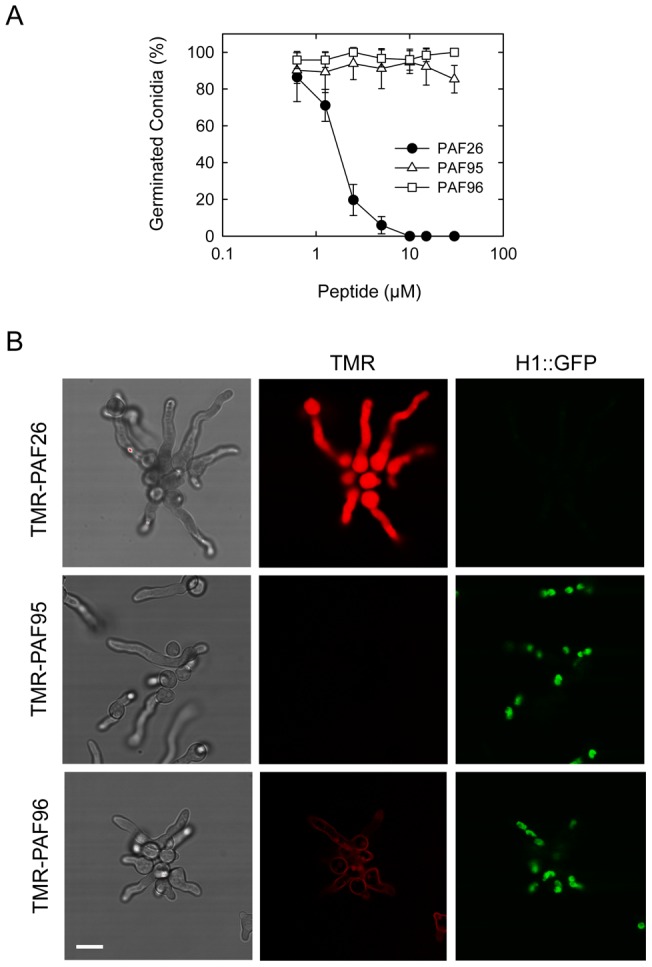
(A) Influence of PAF peptides on conidial germination. Percentage of germinated conidia was quantified by microscopy ∼5 h after treatment with the peptides at different concentrations. (B) Confocal microscopy of the localization of 5 µM TMR-labeled peptides (in red) in germlings of A. fumigatus expressing H1-GFP (in green) 1 h after treatment with the three peptides. Note that the left hand panels show brightfield images of germlings and the right panels show corresponding GFP-labeled nuclei in the same cells. The germlings were all treated for 1 h with the different PAF peptides before imaging. Note that the whole germlings treated with TMR-PAF26 are labeled by the fluorescent peptide, lack nuclei and are dead. This contrasts with both the TMR-PAF95 and TMR-PAF96 treated germlings that possess nuclei and are alive. The TMR-PAF95 treated germlings lack any labeling with the fluorescent peptide whilst TMR-PAF96 is bound only to the cell envelope of the germlings. Bar: 10 µm.