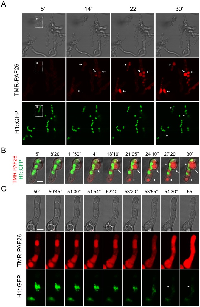
Figure 5. Interaction and internalization of TMR-PAF26 in A. fumigatus.
(A) Sequential images show the interaction and internalization of 5 µM TMR-PAF26 (in red) in actively growing conidial germlings of A. fumigatus expressing H1-GFP (in green) over a period of 25 min. (B) Detail of a germling present in (A) showing merged fluorescent and brightfield images. Note accumulation of TMR-PAF26 in vacuolar compartments (arrows) which undergo expansion and become more intensely fluorescent with time. This coincided with the initiation of the breakdown and loss of fluorescence of the nuclei (asterisks). (C) Detailed images of a germling undergoing these effects over a period of 5 min recorded 50 min after peptide addition. See also Movie S1 for the entire sequential time course of panel (A). Bar: 10 µm in (A) and 4 µm in (B) and (C).

