
Figure 6. Activity and localization of PAF peptides in yeast.
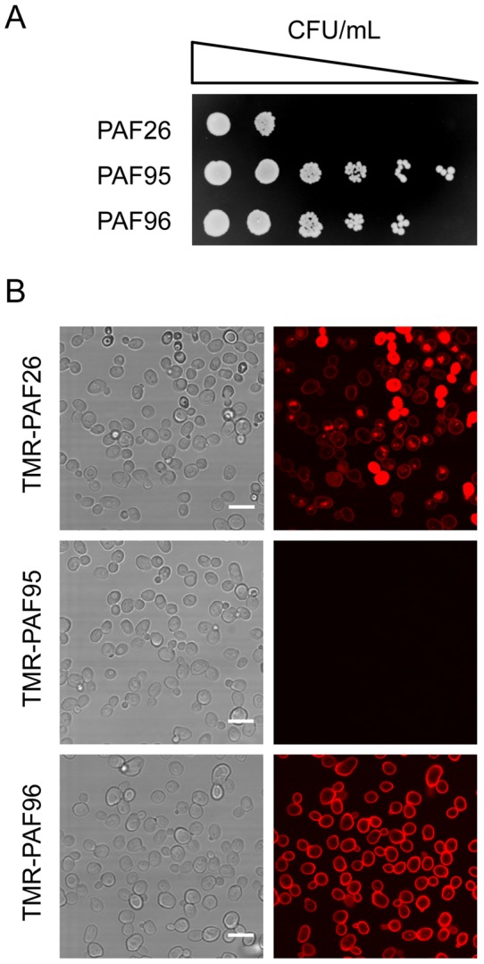
(A) Serial 5-fold dilutions of exponentially growing S. cerevisiae BY4741 cells treated with 64 µM of each peptide for 24 h, and subsequently plated onto YPD peptide-free plates. (B) Brightfield (images on the left) and corresponding confocal microscopy (images on the right) showing the localization in yeast cells (1×10⁶ cells/ml) after treatment with 2.5 µM of each of the TMR-labeled peptides (in red) for 30 min. Bar: 10 µm.