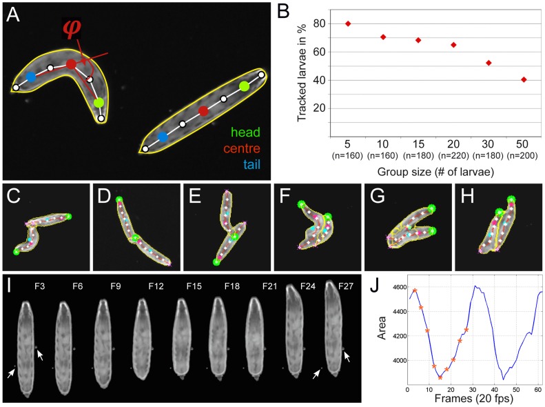
Figure 3. FIM-imaging allows extraction of several features.
(A) Two larvae are shown in the 170 pixel per larval length resolution. The different features extracted are indicated. The spine is indicated by a white line. On the spine seven landmarks are positioned. The head (green dot), the center (red dot) and the tail (blue dot) are indicated. The contour line (yellow) and the bending angle φ are shown. (B) Plot of different group sizes against the percentage of completely tracked paths. The number of larvae tracked (n) is indicated. Recording for 3 minutes at 10 fps and 40 pixel larval length resolution. (C–H) Examples of larval collisions. (C–E) Moderately touching larvae can be separated. (F–H) In case of more intensive collision, the separation of larval contours is not perfect. (I) Stills of a movie showing a larval contraction wave. The numbers indicate the frames shown in (J). (J) The area covered by a larvae changes during contraction. Asterisks indicate the positions of the images shown in (I).

