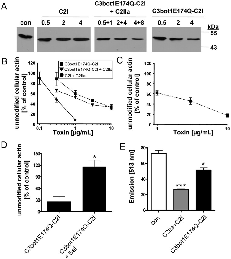
Figure 3. C3bot1E174Q-C2I ADP-ribosylates actin in the cytosol of intact J774A.1 and RAW264.7 macrophages.
A. J774A.1 cells were incubated with C3bot1E174Q-C2I (0.5 µg/mL, 2 µg/mL, 4 µg/mL), C3bot1E174Q-C2I+C2IIa (0.5 µg/mL+1 µg/mL, 2 µg/mL+4 µg/mL, 4 µg/mL+8 µg/mL), C2I alone (0.5 µg/mL, 2 µg/mL, 4 µg/mL) or left untreated. Cells were lysed and lysates incubated for 30 min at 37°C with C2I (300 ng) and biotin-labelled NAD+ (10 µM) to ADP-ribosylate actin, which was not ADP-ribosylated by the toxins in the intact cells. Samples were subjected to SDS-PAGE, blotted and biotinylated (i.e. ADP-ribosylated) actin was detected with streptavidin-peroxidase. Comparable amounts of total protein in the lanes were confirmed by Ponceau S staining (shown in Figure S3). B. Concentration-dependent intoxication of J774A.1 cells with C3bot1E174Q-C2I, C3bot1E174Q-C2I+C2IIa or C2I+C2IIa. Cells were incubated for 6 h with 0.1, 0.3, 1, 3 or 10 µg/mL of the respective or left untreated for control. Subsequently, cells were lysed and further treated as described in A. Intensity of the bands showing ADP-ribosylated actin was determined by densitometry and is given as percentage of actin from untreated control cells (mean±S.D.; n = 3). Comparable protein loading was confirmed by Ponceau S staining (not shown). C. Concentration-dependent intoxication of RAW264.7 cells with C3bot1E174Q-C2I. Cells were incubated for 6 h with 0.1, 0.3, 1, 3 or 10 µg/mL C3bot1E174Q-C2I or left untreated for control. Cells were lysed and treated as described in B. Intensity of ADP-ribosylated actin was determined by densitometry and is given as percentage of actin from untreated control cells (mean±S.D.; n = 3). Comparable protein loading was confirmed by Ponceau S staining (not shown). D. Effect of bafilomycin A1 on intoxication of J774A.1 cells. Cells were pre-treated with 300 nM bafilomycin A1 (Baf) at 37°C and after 30 min C3bot1E174Q-C2I (3 µg/mL) or for control C2I (200 ng/mL)+C2IIa (400 ng/mL) was added to the medium. For control cells were left untreated. Cells were incubated for further 6 h, lysed and further treated as described in A. Intensity of the bands showing ADP-ribosylated actin was determined by densitometry and is given as percentage of actin from untreated control cells (mean±S.D.; n = 3; * = p≤0.5). E. F-actin staining of RAW.264.7 cells treated with C2 toxin or C3bot1E174Q-C2I. Cells seeded in a 96 well plate were treated for 24 h with either C2 toxin as a control (400 ng/mL C2IIa+200 ng/mL C2I) or with C3bot1E174Q-C2I (4 µg/mL) or left untreated. Cells were fixed, permeabilized and F-actin was stained with phalloidin-Alexa-591 and fluorescence detected at 513 nm with an ELISA reader (mean±S.D.; n = 3; * = p≤0.5, *** = p≤0.005).

