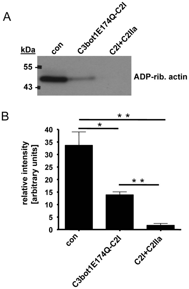
Figure 7. Effect of C3bot1E174Q-C2I on primary cultured macrophages.

Monocytes from human blood were differentiated for 7 days. The resulting macrophages were treated for 6 h at 37°C with C3bot1E174Q-C2I (4 µg/mL) or for control with C2I+C2IIa (200+400 ng/mL) or left untreated. Cells were lysed and lysates incubated for 30 min at 37°C with C2I (300 ng) and biotin-labelled NAD+ (10 µM) to ADP-ribosylate unmodified actin. Comparable amounts of lysate protein were confirmed by SDS-PAGE and Coomassie staining (not shown). The biotinylated (i.e. ADP-ribosylated) actin was detected by Western blotting with streptavidin-peroxidase. B. Intensity of the bands showing ADP-ribosylated actin was determined by densitometry and is given as percentage of actin from untreated control cells (mean±S.D.; n = 3; * = p≤0.5, ** = p≤0.05). B. Comparable amounts of total protein in the lanes were confirmed by Ponceau S staining and anti-Hsp90 Western blotting. B. Morphology of the cells described in A after 6 h. C. HeLa cells were incubated with C3bot1E174Q-C2I (4 µg/mL)+C2IIa (8 µg/mL) or with C3bot1E174Q-C2I alone (4 µg/mL). For control cells were left untreated or were incubated with C2I alone (4 µg/mL). After 6 h of incubation at 37°C all cells were washed, incubated with an antibody against C2I for 5 min at 4°C to remove non-internalized C2I and C2I fusions, washed again and pictures were taken.
