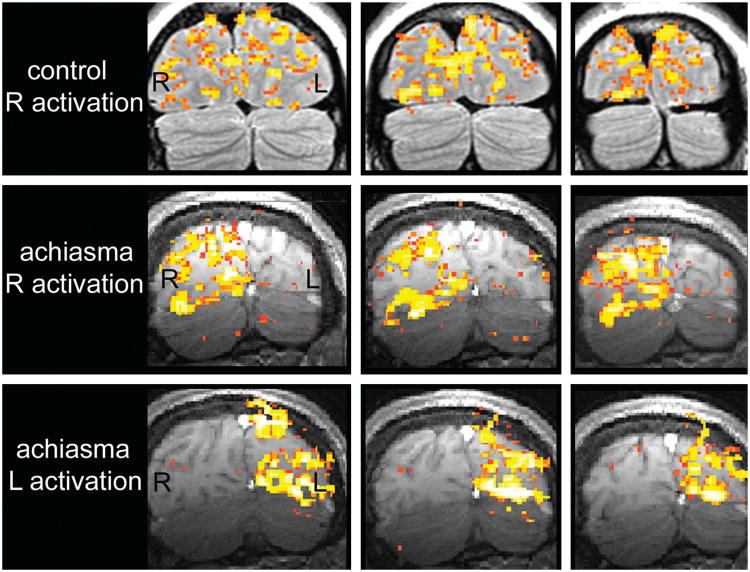
Fig. 3.
Functional MRI during monocular pattern-reversal checkerboard presentation. Coronal images through the visual cortex during right eye stimulation of a control subject (top), during right eye stimulation of our achiasmatic patient (middle), and during left eye stimulation of our achiasmatic patient (bottom). Right eye stimulation of the control subject activates both occipital lobes. Right eye stimulation of the achiasmatic patient activates only the right occipital lobe. Left eye stimulation of the achiasmatic patient activates only the left occipital lobe. These findings are consistent with non-crossing of retinal axonal fibers at the optic chiasm. R, right; L, left.

