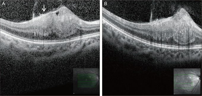
Fig. 2.
Spectral-domain optical coherence tomography reveals an elevated hyperreflective mass in the retina with mild attenuation of the retinal pigment epithelium and photoreceptor inner segment/outer segment junction. Prominent thickening and attenuation of the inner retina is also noted (A,B). The arrow in (A) represents the hyperreflective epiretinal membrane.

