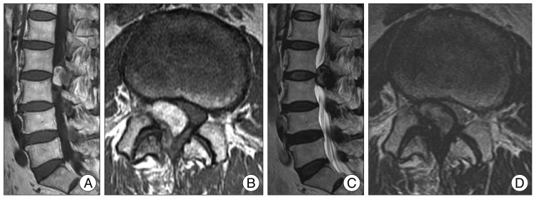
Fig. 1.
Magnetic resonance images are showing the presence of a cystic mass in the right L2-L3 facet joint with arthritis compressing the L3 right root and the dural sac. There are showing a hyperintense on T1-weighted image (A and B) and hypointense on T2-weighted image (C and D) consistent with hemorrhage.

