Fig. 2.

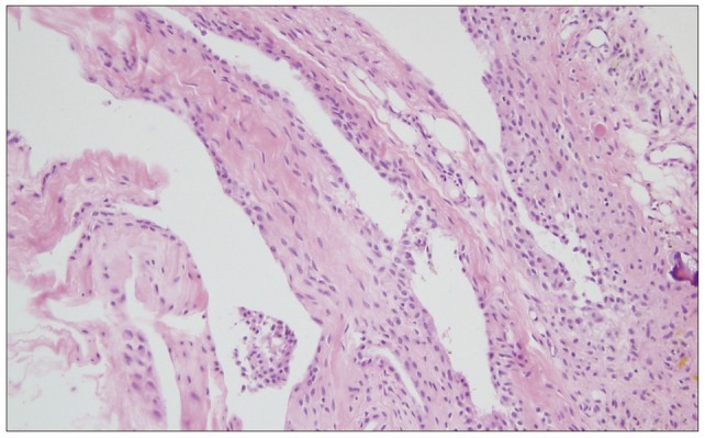
Histological appearance of the hemorrhagic synovial cyst showing synovial cell lining, fibroconnective tissue with widespread hemorrhage, neoangiogenesis, and hemosiderin microdeposits (hematoxylin-eosin stain, ×200).

Histological appearance of the hemorrhagic synovial cyst showing synovial cell lining, fibroconnective tissue with widespread hemorrhage, neoangiogenesis, and hemosiderin microdeposits (hematoxylin-eosin stain, ×200).