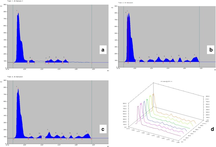
Fig. 4.
Graphical representation of active principle present in callus tissue derived from different PGR containing media as analyzed by CAMAG TLC Scanner-3, Win CATS software version 1.2.3. a Friable callus derived from 2,4-D and KN containing media b Solid non-regenerating callus tissue derived from BAP and IBA containing media c Solid regenerating callus tissue derived from BAP and IBA containing media d Line graph of previous samples, from left in 1st and 4th lane-friable callus tissue loaded 15 μl and 20 μl respectively, in 2nd and 5th lane- Solid non-regenerating callus tissue loaded 15 μl and 20 μl respectively, in 3rd and 6th lane- Solid regenerating callus tissue loaded 15 μl and 20 μl respectively

