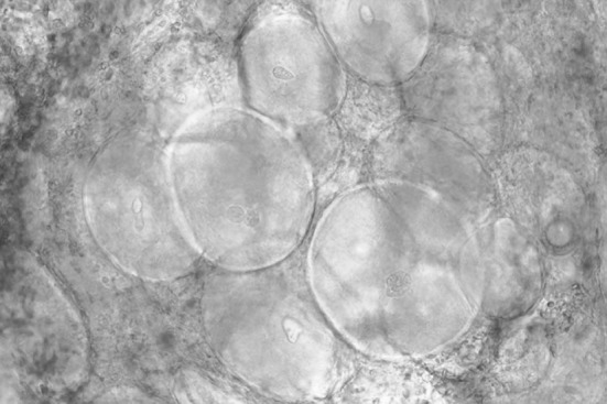
Fig. 3.

A wet mount of tissue from the fin of a largemouth bass (Micropterus salmoides) infected by the lymphocystis virus. The large round cells are epithelial cells containing accumulations of the lymphocystis iridovirus. The diameters of the larger cells are 300–500 μm
