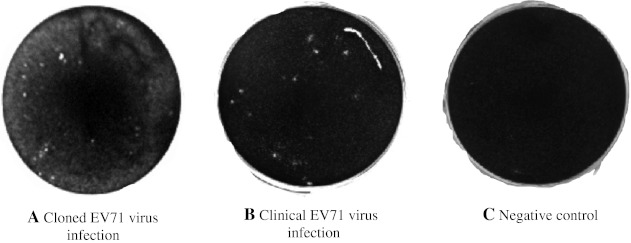
Fig. 5.
Plaque formation of both cloned and clinical EV71: A plaquing assay was performed on both isolates of EV71. The plaques were visualized with crystal violet 3 days post infection in Vero cells. The expected plaques were calculated based on the equation PFU/TCID50 (mL) = 0.7, while the actual number of plaques was counted using the image taken by gel document device (BioRad). The panels show photographs of plaque formation in the wells following infections with: a the cloned EV71 virus, b the clinical EV71 virus, c negative control

