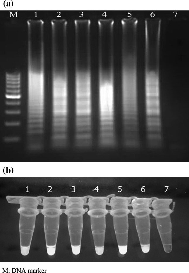
Fig. 2.

(a) Electrophoretic analysis of LAMP amplified products on a 2 % agarose gel; lane M 100-bp DNA ladder marker (Fermentas, Genruler, Germany), lane 1–5 LAMP product of Hepatitis C virus, lane 6 LAMP product of positive control, lane 7 LAMP without target RNA, negative control. (b) SYBR Green I fluorescent dye-mediated monitoring of HCV LAMP amplification. Visual observation of green fluorescence of DNA binding SYBR Green I under UV light, changed to green in the case of positive amplification, whereas in negative control having no amplification, the original orange color is retained; tubes 1–5 LAMP product of HCV, tube 6 LAMP products of positive control, tube 7 LAMP without target RNA. (Color figure online)
