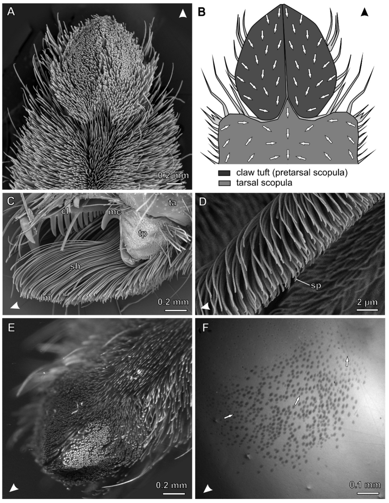
Figure 1. The hairy attachment device of the wandering spider C. salei.
Arrowheads indicate distal direction. (A) SEM micrograph of the distal L1 tarsus. (B) Diagram of the distal part of the tarsus illustrating different scopulae and the orientation of spatula-covered sides of setae (arrows). (C) SEM micrograph of the lateral view of L4 claw tuft, partly shaved. cl, tarsal claw; lm, distal lamellae of setae, bearing the the spatulate setules; mc, middle hook (reduced third claw); sh, seta shafts; ta, tarsus; tp, tenent pad (D) Lateral view of the distal part of a single seta in the median claw tuft. sp, spatula (E) L4 claw tuft of a living C. salei clinging upside-down on a Plexiglas Petri dish. Setae that are in contact are aligned in one plane and reflecting the light of the lateral LED light source at the same angle (bright spots). (F) Same with coaxial illumination (note slightly stronger magnification than in E). Contacting setae appear as darken oval spots, indicating that only the distal tips of setae are in real contact with Plexiglas. Arrows indicate the orientation of setae.

