Fig. 3.
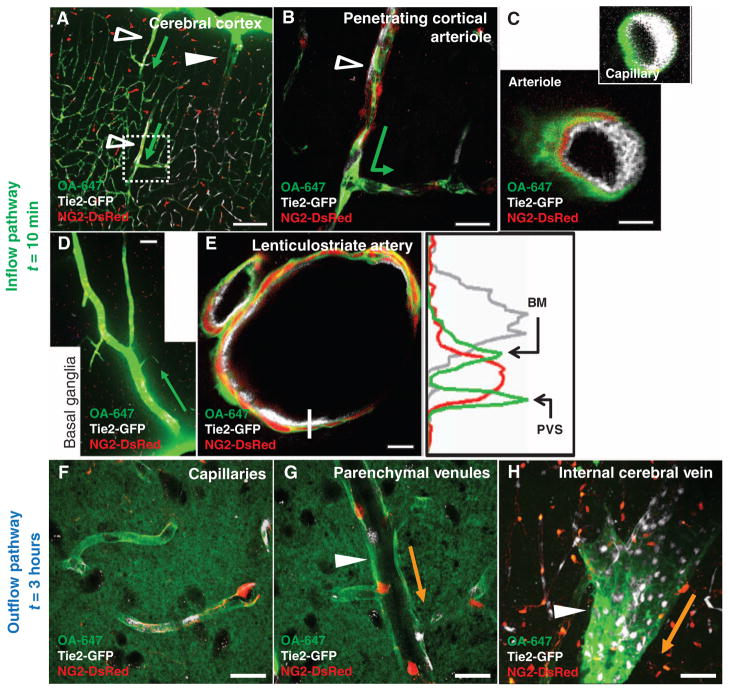
CSF enters and is cleared from the brain interstitium along paravascular pathways. To evaluate the pathways of subarachnoid CSF flux into the brain parenchyma, we injected fluorescent tracer intracisternally into Tie2-GFP:NG2-DsRed double reporter mice, allowing arteries and veins to be directly distinguished. (A and B) Intracisternally injected OA-647 enters (green arrows depict tracer entry) the cerebral cortex along penetrating arterioles (Tie2-GFP+/NG2-DsRed+ vessels, empty arrowheads), not along ascending veins (Tie2-GFP+/NG2-DsRed− vessels, filled arrowheads). (C) CSF tracer moves along both paravascular space (PVS) and the basement membrane (BM) between the vascular endothelial and the smooth muscle cell layers. Tracer movement along capillaries proceeds along the basal lamina. (D) Large amounts of tracer are observed in the basal ganglia and thalamus, entering along large ventral perforating arteries. (E) Detailed tracer distribution around lenticulostriate artery. Plot shows intensity projection along white line. Green, tracer; gray, endothelial GFP; red, vascular smooth muscle. (F to H) At longer time points (>1 hour compared to 0 to 30 min for tracer influx), OA-647 (molecular size, 45 kD) entered the interstitial space and accumulated primarily along capillaries (F) and parenchymal venules (G). Accumulation was greatest along medial interior cerebral veins and ventral-lateral caudal rhinal veins (H). Orange arrows in (G) to (H) depict the observed route of interstitial tracer clearance. Scale bars, 100 μm [(A) and (D)], 50 μm (H), 20 μm [(B), (F), and (G)], and 4 μm [(C) and (E)].

