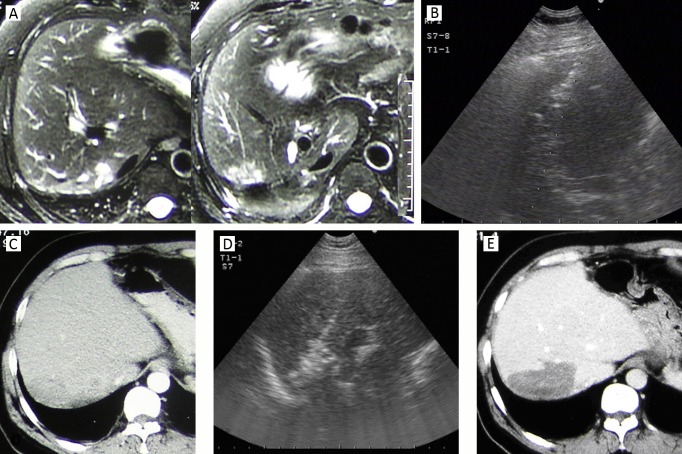
Figure 1.
A 64-year-old man in whom ICC recurred 9 months after partial resection of right hepatic lobe. A. MRI showed an irregular tumor located at the edge of resection on the T2-weighted image; B. Intercostal sonograms showed a 4.2 cm tumor and it was treated by sonography-guided RFA; C. On 1-month CT, the margin of the ablation zone was obscure and coarse, without sufficient safety margin; D. A repeated RFA was performed for the unablated tumor area. E. CT scan 3 months after repeated RFA showed that ablated zone had clear margin and no enhancement. This patient survived for 30 months after the initial RFA treatment and died of tumor spread and metastasis.

